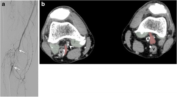
Fig. 4.

Popliteal artery entrapment syndrome (PAES) in a 52-year-old man with a history of right calf claudication for 2 years. (a) Digital subtraction angiography shows abrupt occlusion of the proximal popliteal artery (solid arrow), which reconstitutes at its midportion (dashed arrow). A cine sequence is presented in Movie 1. PAES was suspected and patient underwent CT angiography. (b) Axial images of CT angiography reveal severe narrowing of the right popliteal artery (arrowhead), with an abnormal muscle slip (red transparent overlap) originating from the medial head of the gastrocnemius muscle and coursing between the popliteal vessels (V = popliteal vein). The contralateral side shows a similar appearance of abnormal muscle slip (red transparent overlap); however, the left popliteal artery is patent (A). Bilateral green transparent areas correspond to normal insertions of the gastrocnemius insertions
