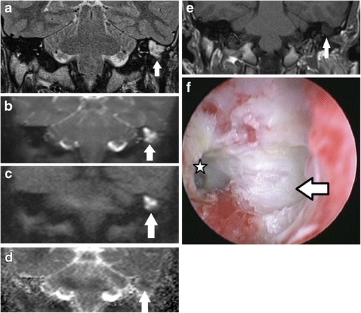
Fig. 1.

Typical MRI characteristics of post-operative cholesteatoma. Coronal images through the left mastoid remnant shows the lesion (white arrow) as high signal on a the T2 weighted image, b DWI b0 image, and c DWI b1000 image and a low signal on the d ADC map and e non-contrast enhanced T1 weighted image. f Surgery confirms the presence of the cholesteatoma. The keratin has been suctioned away showing the underlying white squamous epithelium lining the mastoid remnant (white arrow) and aditus (star)
