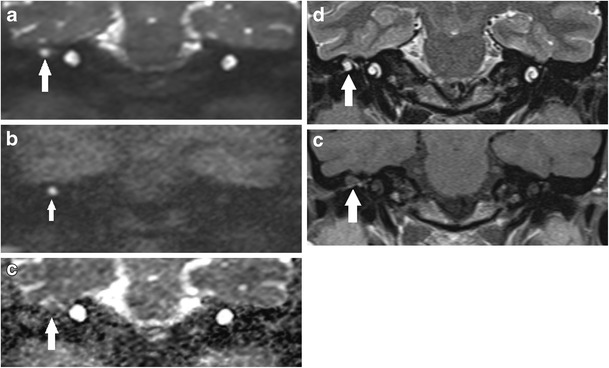
Fig. 3.

DWI appearance of a small residual cholesteatoma confirmed on second-look surgery. The a b0 and b b1000 image shows a small 5 mm high signal cholesteatoma (white arrow) at the right epitympanum. It returns low signal on c the corresponding ADC map, high signal on d the T2 weighted image and low signal on e the non-contrast enhanced T1 weighted image
