
FIGURE 1.
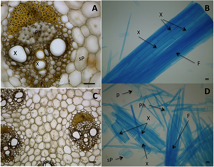
Biomass units of sugarcane stem (culm) with cells bound by middle lamellae. (A,C) Stele, the vascular bundle showing Fibers (F), Phloem (Ph), Xylem (X), sucrose-storage parenchyma (sP). (B) Macerated tissue showing a detail of an isolated stele that still maintains adhesion among cells. (D) Cells of sugarcane stem without the binding of middle lamellae. Cells are named as in (A). The cell separation was prepared according to Franklin (1945) and cells were stained with toluidine blue (B,D) and iodine-potassium iodide (A,C). Bars represent 100 μm.