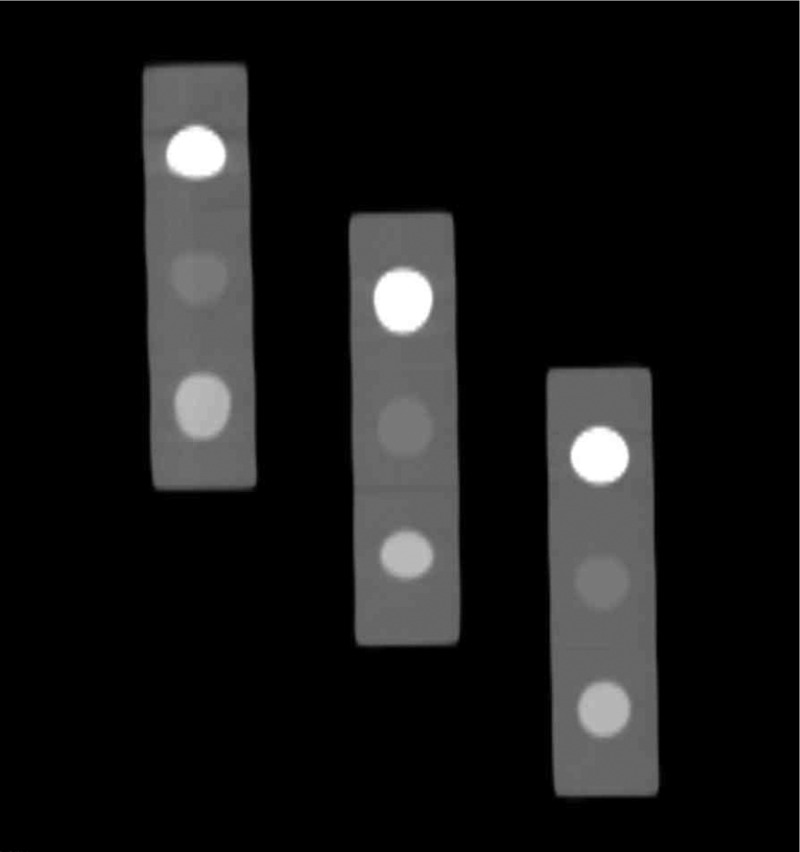
Fig. 5.
A CT scan performed in the United States of three second-generation CT pocket phantoms using the same model CT scanner observed to have the highest variability in the clinical trial. Slice thickness and spacing for this scan was 1.25 mm. No patient is present in this scan and spatial warping is evident along the direction (top to bottom), particularly for the phantom that is furthest from scanner iso-center (left side).

