. 2016 Sep 8;59(5):505–511. doi: 10.3340/jkns.2016.59.5.505

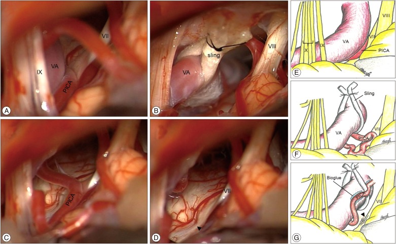
Fig. 2. Intraoperative microscopic view during MVD. A : The loop of the VA and the PICA are seen compressing the root exit zone (REZ) of the facial nerve. B : The VA was adequately lifted using the sling, which was fixed to the 5-0 black silk sutures on the petrosal dura. C : PICA was fully identified on the REZ only after transposition of the VA. D : An indentation was seen on the REZ after the decompression (arrowhead). E : Schematic illustration showing compression of the facial nerve by the VA and the PICA. F : After transposition of the VA using a bioglue-coated Teflon sling, the PICA compressing on the REZ of the facial nerve is fully identified. G : The PICA was fixed to the transposed VA with applications of several pieces of TachoSil (Takeda GmbH, Linz) and Tisseel bioglue (Baxter). MVD : microvascular decompression, VA : vertebral artery, PICA : posterior inferior cerebellar artery.

Fig. 2