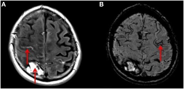
Figure 3.

(A) T2 FLAIR axial MRI of the brain showing right-sided ICH and acute bleeding (red arrows). (B) SWI sequence MRI of the same patient demonstrating a linear gyriform hypointensity in a cortical distribution (red arrow), representative of siderosis, in a region independent to that of the site of hemorrhage.
