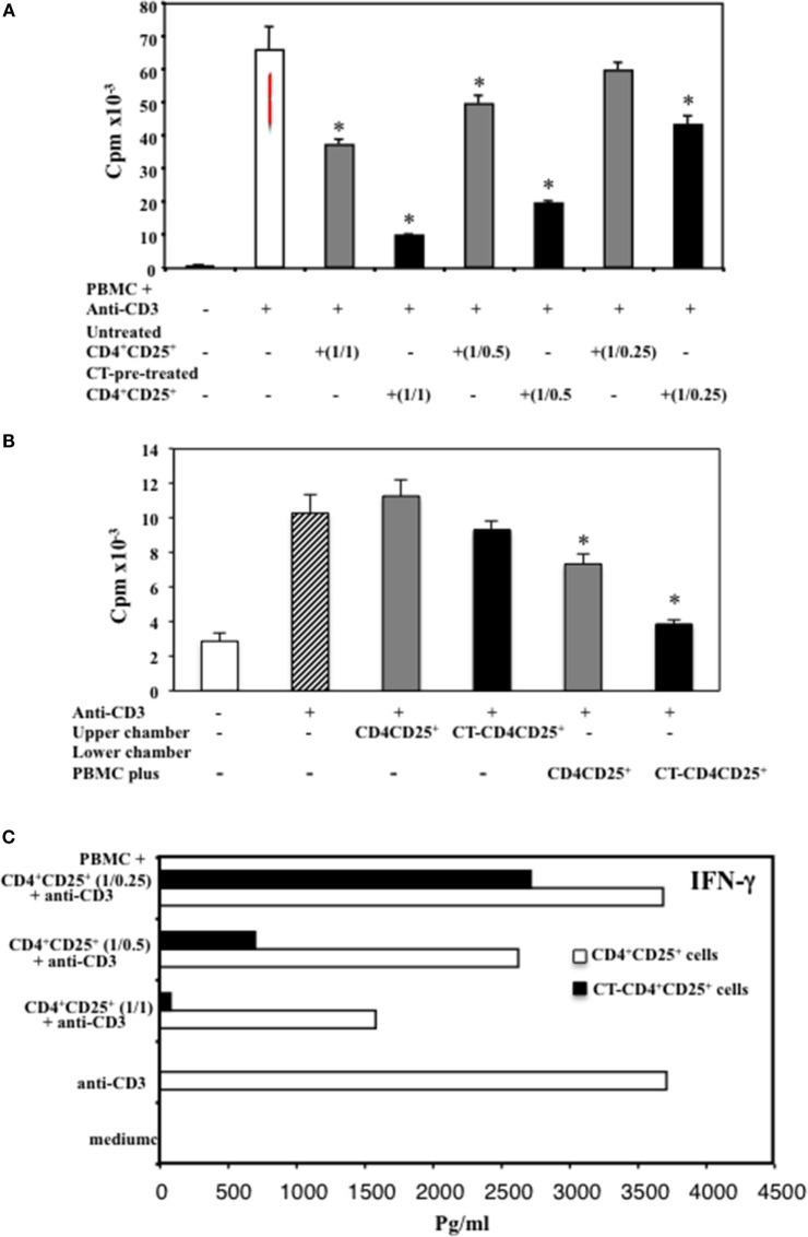
Figure 3.
Enhanced suppression of T cell proliferation and IFNγ inhibition by CD4+CD25+ T lymphocytes pre-treated with CT. (A) Immunomagnetically purified CD4+CD25+ T lymphocytes isolated from healthy donors were cultured in the presence or absence of CT (1 μg/ml). After overnight incubation, cells were washed three times and cultured with autologous PBMC at different ratios (1/1, 1/0.5, 1/0.25) in triplicate in 96-well plates and stimulated with anti-CD3 mAbs. (B) Alternatively, PBMC (1 × 10⁶) were placed in the lower wells of transwell and untreated or CT-pre-treated CD4+CD25+ T lymphocytes (1 × 10⁶) were cultured either in the upper wells or in the lower chambers in contact with PBMC. Cells were stimulated with anti-CD3 mAb (0.5 μg/ml), and the PBMC proliferation was evaluated after 66 h by 3H-thymidine incorporation. The bars indicate the mean of triplicate wells. (C) After 48 h, the amount of IFNγ was valuated in the supernatants from the different cultures by ELISA assay in duplicate. The * indicate that the differences between CT-treated as compared with untreated cells are significant (p < 0.05). One representative for each of three experiments performed is shown.

