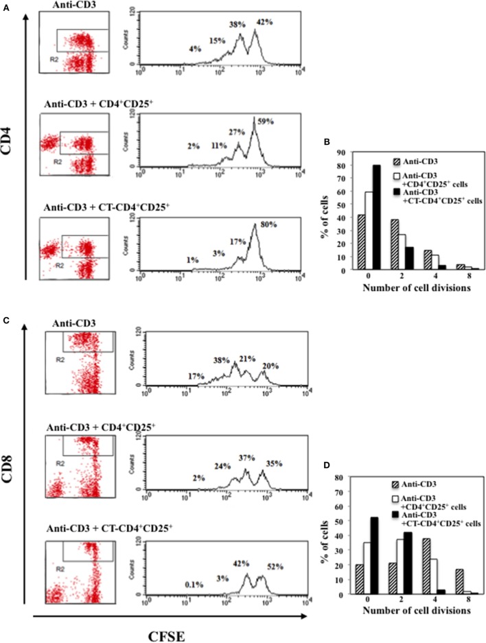
Figure 4.
Inhibitory effects of CD4+CD25+ T lymphocytes pre-treated and untreated with CT on CD4 or CD8 T cell proliferation. Immunomagnetically purified CD4+CD25+ T lymphocytes isolated from healthy donors were cultured in the presence or absence of CT (1 μg/ml). After overnight incubation, cells were washed three times and cultured with autologous CFSE-labeled PBMC at 1/1 ratio and stimulated with anti-CD3 mAbs. The percentage of proliferating CD4+ (A,B) and CD8+ (C,D) T cells was evaluated by FACS analysis after 5 days of culture by CFSE dilution on CD4 and on CD8-gated populations. The numbers within the plots and the graphs indicate the percentage of cells that went through 0, 2, 4, and 8 cell divisions. One representative of three performed experiments is shown.

