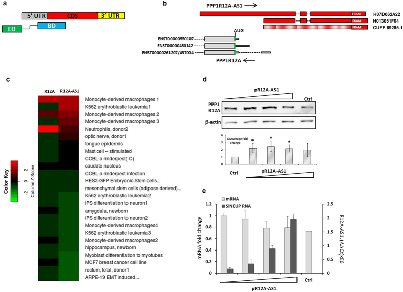
Figure 1. Expression and functional characterization of R12A-AS1.
(a) Schematic overview of a miniSINEUP and the target mRNA. 5′ and 3′ UTRs, coding sequence (CDS) of the mRNA and the Binding Domain (BD) and Effector Domain (ED) of the SINEUP are indicated. BD and ED are connected with a spacer sequence, (shown as a black elbow line). (b) Genomic organization of the PPP1R12A sense-antisense overlapping region (the elements are not in scale). PPP1R12A coding exons are shown as thick grey bars, UTRs as thin grey bars. R12A-AS1 exons in the two RIKEN full-length cDNA clones are shown in red, and introns by dashed lines. Green vertical lines indicate the position of the AUG codon in the mRNA. The R12A-AS1 isoform, assembled by Cufflinks, is shown by a patterned bar, and the position of the FRAM element is indicated by a red box. (c) Expression of PPP1R12A and R12A-AS1 in the FANTOM5 dataset. The tpm values for the top 25 samples of Supplementary Table 3 are presented as a matrix plot. (d) Western blot analysis of PPP1R12A in HEK293T cells transfected with 30, 60, 150 and 300 pmol of pR12A-AS1, or empty vector as control, respectively. β-actin is shown as a loading control. Lower panel shows mean intensity of PPP1R12A bands, normalized to actin B. (e) Corresponding RNA levels, detected by RT-qP PCR (mean ± s.d. n = 3). *(n = 3, mean + S.D., p < 0.05, One-Sample t-Test, vs empty vector).

