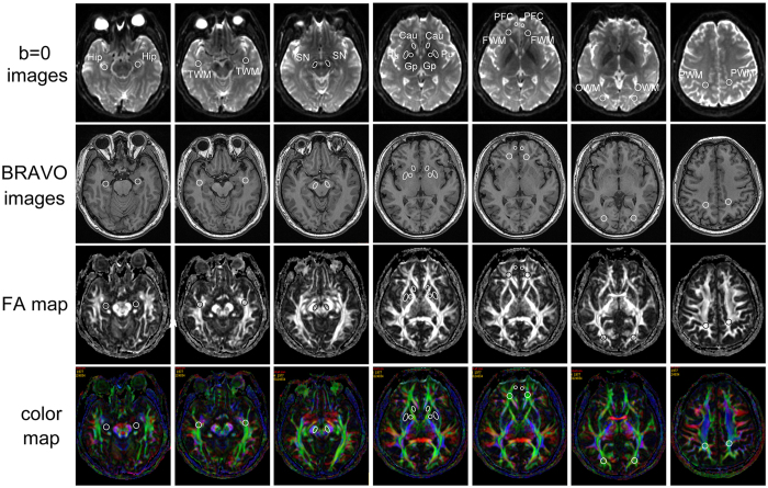
Figure 4. Region of interest (ROI) settings.
Cau, caudate nucleus; FWM, frontal white matter; Gp, globus pallidus; Hip, hippocampus; OWM, occipital white matter; PFC, prefrontal cortex; PWM, parietal white matter; Pu, putamen; SN, substantia nigra; TWM, temporal white matter. The individual regions are drawn on DTI MR images (4,600/82.9) on the fractional anisotropy (FA) map, color map, b = 0 image and BRAVO map (8.2/3.2).

