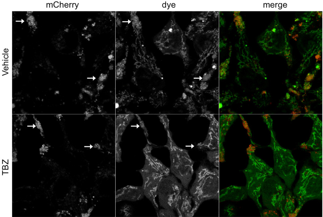
Figure 1. Images acquired on Nikon A1R laser scanning confocal.
Images acquired with Nikon A1R laser scanning confocal. The high resolution of this microscope allows for the identification of the punctate localization of the dye in mCherry-positive structures (indicated by arrows) that are localized mostly within extensions from the cell body. Furthermore the mitochondrial staining pattern is also visible. When VMAT2 function is inhibited by TBZ, the difference in staining is easily observed. Dye can only be seen in the mitochondrial compartment; punctate staining within the mCherry-positive puncta is lost.

