
Figure 3.
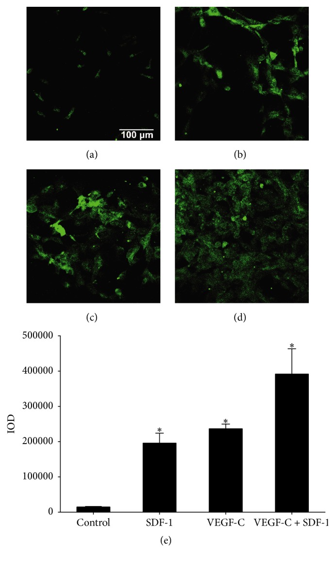
LECs with positive LYVE-1 immunostaining differentiated from CD34+/VEGFR-3+ EPCs. The CD34+/VEGFR-3+ cells were cultured in the presence of SDF-1 (100 ng/mL) and VEGF-C (60 ng/mL). The cells expressed LYVE-1 marker after induction with no addition ((a), control), SDF-1 (b), VEGF-C (c), and SDF-1 + VEGF-C (d) for two weeks. Integral optical density (IOD) of LYVE-1 fluorescence of each group (e). Values are expressed as mean ± SD, n = 3. ∗ p < 0.01, significant increase versus control group.