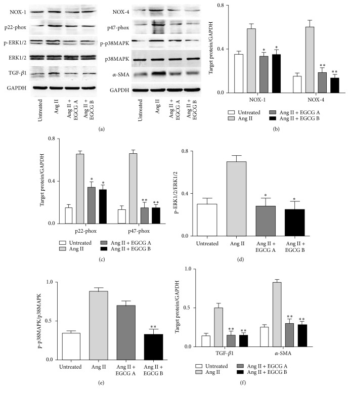
Figure 6.
The protein expression of NOX-1 and NOX-4 (b), p22-phox and p47-phox (c), p-ERK1/2 (d), p-P38 MAPK (e), and TGF-β1 and α-SMA (f) of HK-2 cell after exposure to Ang II and EGCG detected by Western blot analyses. HK-2 cells were pretreated with 15 or 30 μM of EGCG for 6 h and then stimulated with Ang II (1 μM) for 24 hours. Values are means ± SEM. ∗ P < 0.05 and ∗∗ P < 0.01 versus control.

