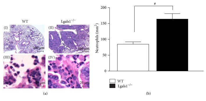
Figure 3.
Lgals1−/− mice have increased neutrophil infiltration in the lung parenchyma. H. capsulatum-infected mice were euthanized on day 15 after infection and lung sections (5 μm) were embedded in paraffin blocks. Lung sections from WT + H. capsulatum (I, bar: 100 μm; III, bar: 25 μm) and Lgals1−/− + H. capsulatum (II, bar: 100 μm; IV, bar: 25 μm) were stained with hematoxylin (a) and neutrophils were quantified (neutrophils/mm2) using magnifications ×400 (b). Data are representative of one of the two experiments performed independently (n = 10 per group). Values are mean ± SEM. # p < 0.05 WT versus Lgals1−/−, both infected.

