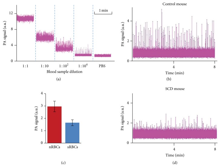
Figure 6.
In vitro PAFC of sRBCs. (a) PA signal amplitudes from nRBCs of SCD mouse, flowing in a capillary tube at different dilutions showing completely overlapped (1 : 1) and nonoverlapped (1 : 104) signal peaks from individual sRBCs. (b, d) Typical PA signal traces with nonoverlapping peaks from individual nRBCs of a nude mouse (b) and sRBCs of a SCD mouse (d). (c) Averaged PA signal amplitudes from nRBCs and sRBCs in (b, d).

