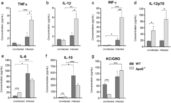
Figure 7. ApoE−/− mice have modulated levels of serum pro-inflammatory and anti-inflammatory cytokines.
Serum concentrations of (A) TNF-α, uninfected ApoE−/− vs. infected ApoE−/− P = 0.0002, infected WT vs. infected ApoE−/− P = 0.0166; (B) IL-1β, uninfected ApoE−/− vs. infected ApoE−/− P = 0.0064, infected WT vs. infected ApoE−/− P = 0.0349; (C) IFN-γ, uninfected WT vs. infected WT P = 0.0241, uninfected ApoE−/− vs. infected ApoE−/− P = 0.0009, infected WT vs. infected ApoE−/− P = 0.0042; (D) IL-12p70, uninfected WT vs. uninfected ApoE−/− P = 0.0386, infected WT vs. infected ApoE−/− P = 0.0166; (E) IL-6, uninfected WT vs. uninfected ApoE−/− P < 0.0001, uninfected WT vs. infected WT P = 0.0155, uninfected ApoE−/− vs. infected ApoE−/− P = 0.0002; (F) IL-10, uninfected WT vs. uninfected ApoE−/− P = 0.0017, uninfected WT vs. infected WT P = 0.0412, uninfected ApoE−/− vs. infected ApoE−/− P = 0.0002; and (G) KC/GRO, uninfected WT vs. uninfected ApoE−/− P < 0.0001, uninfected ApoE−/− vs. infected ApoE−/− P = 0.0005 from uninfected and infected WT and ApoE−/− mice. n = 5 for all groups. *P ≤ 0.05, **P ≤ 0.01, and ***P ≤ 0.0001. Infected, WT and ApoE−/− mice were analyzed on day 7 to 9 post-infection (upon the development of clinical symptoms in the WT mice).

