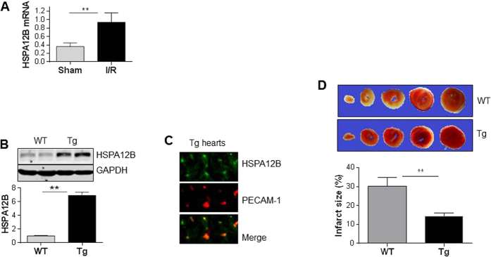
Figure 1. HSPA12B overexpression in endothelial cells limited infarct size following myocardial I/R.
(A) Upregulation of HSPA12B by I/R. Ventricular tissues were collected from WT mice 4 h after I/R. Total RNA was extracted for examination of HSPA12B mRNA levels. The mRNA levels of β-actin served as internal controls. **P < 0.01, n = 6 per group. (B) Overexpression of HSPA12B in Tg hearts. Ventricular tissues were collected from 8-week old mice. Cellular extracts were prepared for immunoblotting against HSPA12B. The blots against GAPDH served as loading controls. **P < 0.01, n = 4 per group. (C) Colocalization of HSPA12B with PECAM-1 in Tg hearts. Ventricular tissues were collected from HSPA12B Tg mice aged of 8-week old. Cryosectioning was prepared for the immunofluorescence staining against HSPA12B and PECAM-1, a selective marker of endothelial cells. Note that the staining of HSPA12B (green) was colocalized with PECAM-1 (red). The representative images were from three independent mice. (D) Infarct size. TTC staining was performed to analyzed infarct size (pale white) 4 h after I/R. **P < 0.01, n = 5 per group.

