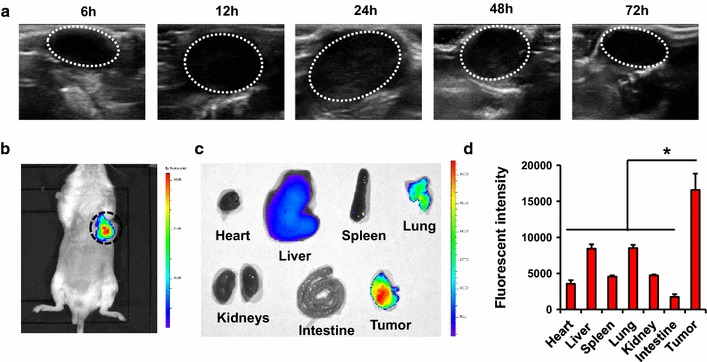
Fig. 3.

Accumulation of the nano-oxygen-carrier in tumor site. a Ultrasound imaging of the nano-oxygen carrier in tumor-bearing mice after intravenous injection of lip(PFH). Images were taken at 6–72 h post-injection. Tumors were circled by white dash line. b Near-infrared imaging of IR780 in tumor-bearing mice after intravenous injection of lip(IR780 + PFH). Imagines were taken 24 h post-injection. Tumors were circled by blue dash line. c Near-infrared imaging of IR780 in the different organs from tumor-bearing mice 24 h post-injection of lip(IR780 + PFH). d Quantitation of near-infrared signals in the organs from tumor-bearing mice 24 h post-injection of lip(IR780 + PFH). *P < 0.05 compared with tumor
