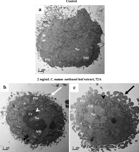
Fig. 5.

Transmission electron micrographs of the control (untreated) D24 cell (a) and cells treated with the crude MeOH extract (2 mg/mL) for 72 h (b & c). Distinct morphological changes, including plasma membrane alteration (white arrow), chromatin condensation (white arrowhead), blebbing (black arrow) and segmented/lobulated nucleus (black arrowhead) were observed in the treated cells. Cr: chromatin, MV: microvilli, NM: nuclear membrane, Nu: nucleus, PM: plasma membrane
