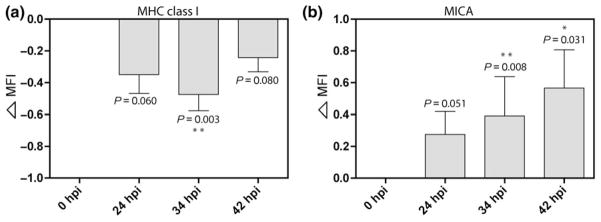
Fig. 1.
Alterations in MHC class I and MICA expression on A2EN endocervical epithelial cells after Chlamydia trachomatis infection. Flow cytometric assessment of MHC class I and MICA expression on mock-infected cells and C. trachomatis-infected A2EN cells at 24, 34, and 42 hours postinfection (hpi). Chlamydia trachomatis-infected cells were gated based on Chlamydial-LPS-FITC positivity. Data shown are representative of means and standard deviations of ‘delta MFI’ from four independent experiments. (a) MHC class I expression of C. trachomatis-infected cells was significantly downregulated at 34 hpi (**P < 0.01). (b) MICA expression progressively increased over time postinfection. Statistical analyses were performed using Student’s t-test; P-values are included to demonstrate the trends toward significant differences in MHC class I and MICA expression of A2EN cells at different times post-C. trachomatis infection when compared to the mock-infected control. The formula for calculating ‘delta MFI’ is described in materials and methods; a delta MFI of 0.5 represents a 100% increase in the absolute MFI value relative to mock-infected cell ligand expression.

