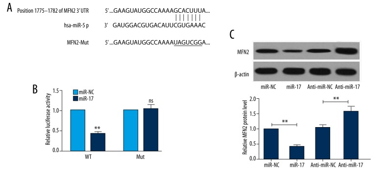
Figure 4.
MFN2 is a target of miR-17. (A) Wild-type (WT) and mutant (Mut) 3′UTR binding sites are shown. The mutated bases are labeled with a horizontal line. (B) HEK293 cells were co-transfected with WT or Mut MFN2 3′-UTR and miR-17 mimic or miR-NC. The relative luciferase activity was detected 48 h after transfection. (C) At 48 h post-transfection, Western blot analysis was performed to assess the effects of miR-17 and aiti-miR-17 on expression of MFN2 protein. Data are expressed as the mean ±SD (n=3). * P<0.01, ** P<0.01; NS – no significant difference compared to control.

