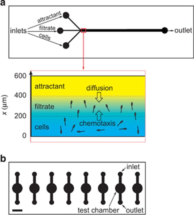
Figure 1.
Schematics of the microfluidic devices used to study the temperature dependence of motility behaviors of the coral pathogen Vibrio coralliilyticus. Both panels presented from the microscopist's perspective, where the objective is directly below the image shown and the devices are mounted onto a standard 1 × 3 inch microscope slide. (a) Planar view of the three-inlet channel used for the chemotaxis experiments. Three inlets converge into a 600 μm-wide, 100 μm-deep channel, creating three, 200 μm wide streams of cells, filtrate and attractant, respectively. The imaging window is denoted by the red box. (Inset) Diffusion of the attractant across the channel (x direction) creates chemical gradients to which bacteria can respond by chemotaxis. (b) Parallel holding chambers, 100 μm in depth and 5 mm in diameter, used for the chemokinesis experiments. Bacteria injected through the inlet were observed in the test chamber under quiescent conditions. The eight chambers on a chip allowed for parallel experiments in conjunction with the automated, programmable microscope stage that can image successive chambers efficiently and return to the precise imagine position each time. Scale bar=5 mm.

