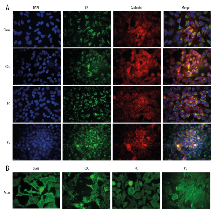
Figure 1.
Immunofluorescent staining of HeLa cells cultured and stained on four different materials. HeLa cells (ATCC, Manassas, Virginia, USA) were cultured overnight either on glass coverslips or on the apical compartment of 12 mm Transwell® inserts (Corning Inc.; Corning, New York, USA) composed of three membrane materials: collagen-coated polytetrafluoroethylene (COL), polycarbonate (PC), and polyester (PE). (A) Cells stained with Calnexin monoclonal antibody (endoplasmic reticulum marker) and rabbit polyclonal to pan Cadherin (cell membrane marker). (B) Cells stained with monoclonal anti-β-actin-FITC conjugate clone AC-15.

