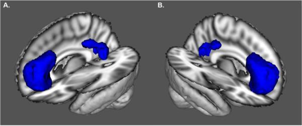
Figure 1. Default Mode Network Region of Interest.
Anterolateral view of the left (A) and right (B) default mode network (DMN) region of interest (ROI). The ROI was composed of the ventral medial prefrontal cortex and posterior cingulate/precuneus. The DMN ROI was identified using an Independent Component Analysis (ICA). Cerebral blood flow (CBF) was quantified within this ROI using arterial spinal labeling (ASL) MRI. The underlay represents the 3D reconstruction of the MNI152 T1-weighted 2 mm brain.

