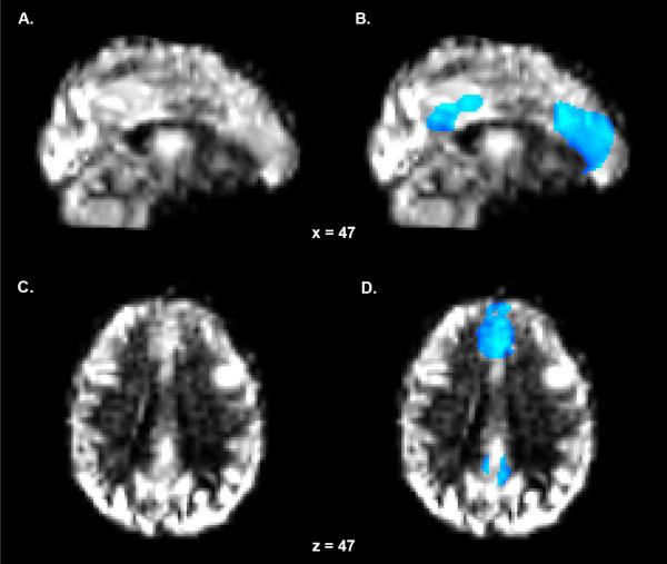
Figure 2. Perfusion Map and DMN Region of Interest Mask.
(A) Sagittal and (C) axial perfusion calibrated cerebral blood flow (CBF) maps of a single representative subject. The CBF maps were warped to standard space using the non-linear matrix generated during the transformation of the anatomical volume to standard space. (B) Sagittal and (D) axial representations of the DMN region of interest mask overlaid on top of the CBF map. The mask is scaled to CBF (blue-light blue) and thresholded at 15.

