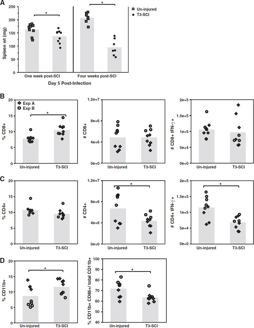
Fig. 3.
Spleen size and the generation of an adaptive immune response are reduced following infection of T3–injured mice. Spleen size increases following infection, thus on day 5 p.i. with 1 × 10⁴ PFU MHV spleen weight and cellular composition were assessed. A) Following infection at 1 week and 4 weeks post-SCI, T3-injured mice showed significantly reduced spleen weight compared to un-injured mice (*p ≤ 0.03). B–D) At 1 week post-SCI, T3-SCI mice were infected with 1 × 10⁴ PFU and the generation of an adaptive immune response in the spleen evaluated on day 5 p.i. using flow cytometric analysis. Virus-specific T cells were determine by ex vivo stimulation with CD4 Ag-specific peptide (epitope M133–147), CD8 Ag-specific peptide (epitope S598–605), or non-specific OVA control peptide, prior to flow cytometric assessment. T3-injured mice showed significant increase (*p ≤ 0.002) in the frequency of CD8+ T cells compared to un-injured mice (B, first panel), however there was no difference in the number of total CD8+ T cells nor the number of virus-specific CD8+ IFN-γ+ T cells (B, second and right-hand panels). The frequency of CD4+ T lymphocytes was similar between infected T3-injured and un-injured mice (C, first panel), however the numbers of total CD4+ and virus-specific CD4+ IFN-γ+ T cells were significantly reduced in injured mice (*p ≤ 0.04) (C, second and right-hand panels). The frequency of CD11b+ macrophages was significantly increased in T3-injured mice (*p ≤ 0.04) (D, first panel), however the frequency of CD86+ activated CD11b+ macrophages within the total macrophage population was significantly decreased compared to un-injured mice (*p ≤ 0.01) (D, second panel). The mean number of cells is presented in bar graphs, with each data point representing one mouse from two separate experiments.

