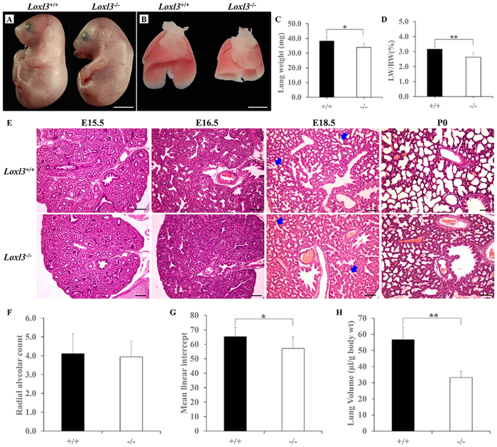
Figure 1. Lung defects in LOXL3 knockout mice.
(A) Loxl3−/− mice exhibited spinal abnormalities at E18.5. Bar: 5 mm. (B) The Loxl3−/− mouse lungs were smaller in size than the Loxl3+/+ mouse lungs at E18.5. Bar: 2 mm. (C,D) Lung weights and the LW/BW ratios of the Loxl3−/− mice were significantly decreased compared with the Loxl3+/+ mice at E18.5. *P < 0.05. **P < 0.01. (E) No significant differences appeared in the lung structures between Loxl3−/− and Loxl3+/+ mice until E15.5. The terminal lung tubules (acini) of the Loxl3−/− mice did not dilate at E16.5. A significant reduction in the distal saccular spaces in Loxl3−/− lungs was noted from E18.5 onwards. Blue arrows: distal saccules. Bar: 100 μm. (F) Normal RACs were observed in the Loxl3−/− mouse lungs at E18.5. (G) Decreased MLIs were observed in the Loxl3−/− mouse lungs at E18.5. *P < 0.05. (H) The lung volumes of the Loxl3−/− mice were also significantly decreased at E18.5. **P < 0.01. For all bar graphs, n = 5 in each group.

