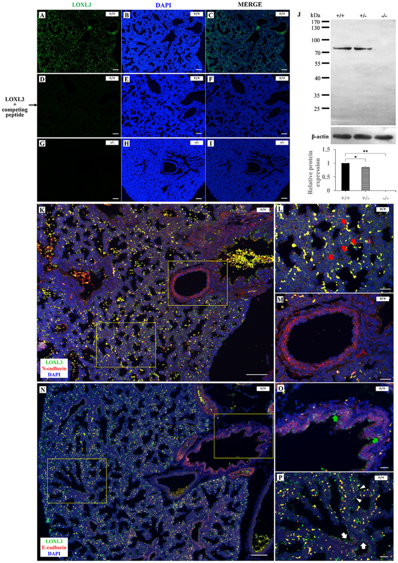
Figure 3. LOXL3 expression in lungs at E18.5.
(A–C) Immunofluorescence staining indicated that LOXL3 was abundantly expressed in wild-type mouse lungs. (D–F) No positive signal was detected by the LOXL3 antibody in wild-type mouse lungs in the presence of the LOXL3 competing peptide. (G–I) LOXL3 expression was not detected in LOXL3-deficient mouse lungs. (J) Western blots for LOXL3 and β-actin in Loxl3+/+, Loxl3+/−and Loxl3−/− lungs were analysed at E18.5. *P < 0.05, **P < 0.01. n = 5 in each group. (K) The co-staining of LOXL3 (green) and N-cadherin (red, a mesenchymal marker) in wild-type mouse lungs at E18.5. (L,M) Enlarged views of Fig. K. (L) N-cadherin stained the mesenchyme, and LOXL3 was found to localize primarily in the mesenchyme (red arrows). (M) LOXL3 was rarely expressed in the blood vessels of the lung. (N) The co-staining of LOXL3 (green) and E-cadherin (red, an epithelial marker) in wild-type mouse lungs at E18.5. (O,P) Enlarged views of Fig. N. (O,P) LOXL3 was expressed at lower levels at the interface between the epithelial cells of the proximal airways (green arrows), distal airways (white arrows) and respiratory epithelium (white arrowheads). DAPI was used to stain the nucleus. Bars: 100 μm (A–I,K,N); 25 μm (L,M,O,P).

