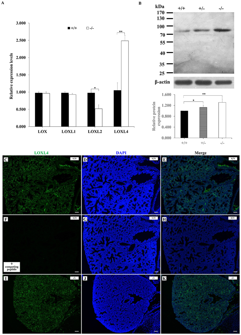
Figure 6. Loxl4 expression in the lung.
(A) The Loxl2 mRNA level was significantly reduced in the Loxl3−/− lungs compared with the Loxl3+/+ lungs. *P < 0.05. Conversely, the Loxl4 mRNA levels in the Loxl3−/− lungs were significantly increased compared with the Loxl3+/+ lungs. **P < 0.01. n = 3 in each group. (B) The LOXL4 protein levels in the lung were analysed by Western blot and normalized to β-actin expression. Loxl3−/− lungs contained higher levels of the LOXL4 protein than Loxl3+/+ lungs. *P < 0.05, **P < 0.01. n = 5 in each group. (C–K) The distribution of LOXL4 in the Loxl3+/+ and Loxl3−/− lungs was analysed by immunofluorescence. (C–E) LOXL4 was found to express widely in the Loxl3+/+ lung. (F–H) LOXL4 expression was not detected in the Loxl3+/+ lung in the presence of the LOXL4 competing peptide. (I–K) The high expression of LOXL4 was also observed in the Loxl3−/− lungs. DAPI was used to stain the nucleus. Bar: 100 μm.

