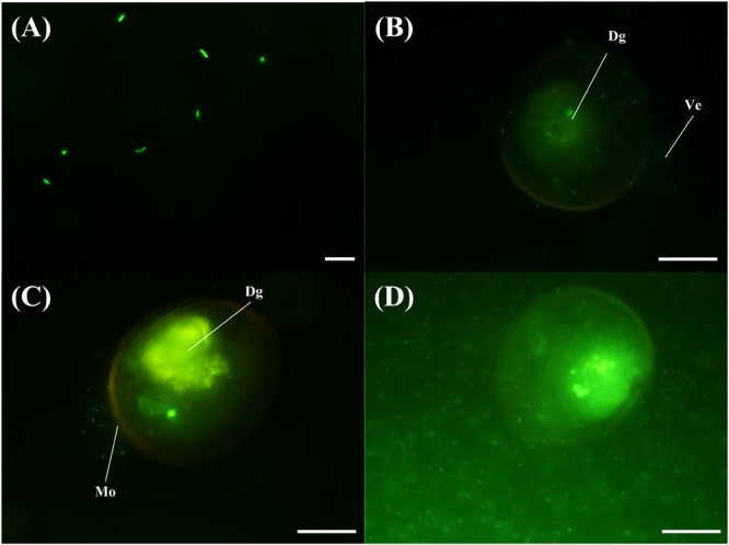
FIGURE 5.

Invasive activity of V. tubiashii VPAP30 strain on larvae of scallop A. purpuratus determined by epifluorescence microscopy. Bacterial cells of V. tubiashii stained with 5-DTAF (A); Stained V. tubiashii in the digestive gland at 30 min post-infection (B); Bacterial cells of V. tubiashii in the digestive gland at 1 h post-infection (C); Bacterial cells invading completely the larval body cavity and surrounding shell at 24 h post-infection. (Dg) Digestive gland; (Ve) Velum; (Mo) Mouth. Scale bars: 10 μm (A); 50 μm (B-D).
