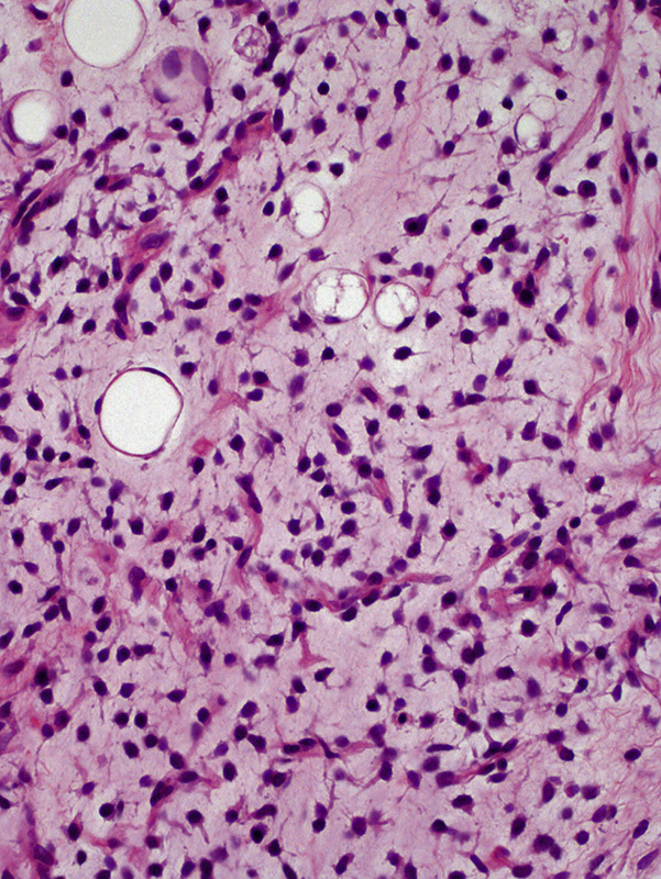
FIGURE 2.

Primary retroperitoneal MLS showed a classic histologic appearance, characterized by a mixture of uniform oval cells and signet-ring cell lipoblasts on a background comprising myxoid stroma and plexiform capillary network (hematoxylin and eosin staining).
