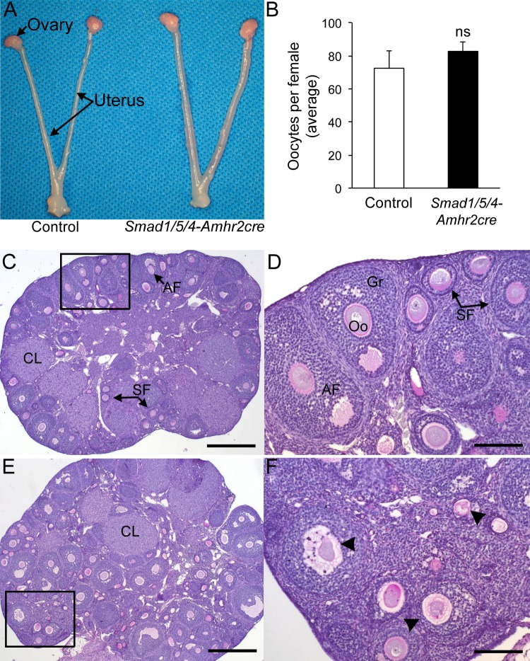
FIG. 1.
Ovarian histology and ovulation in Smad1/5/4-Amhr2-cre KO female mice. A) Reproductive tract of Smad1flox/flox Smad5flox/flox Smad4flox/flox control (left) and Smad1flox/flox Smad5flox/flox Smad4flox/flox Amhr2cre/+ KO mice at 6 wk of age. No gross differences were detected. B) Pharmacologic superovulation of control and Smad1/5/4-Amhr2-cre KO mice at 3 wk compared to control. No statistical differences (ns) were detected in the mean number of oocytes collected in the oviducts of control; n = 3 each genotype. C) PAS stain of an ovary from a 12-wk-old Smad1flox/flox Smad5flox/flox Smad4flox/flox control mouse shows normal follicular development with multiple corpora lutea (CL) and growing follicles (SF, secondary follicles; AF, antral follicle). D) Inset shown in panel C shown at 100x magnification. E) PAS stain of an ovary from a 12-wk-old Smad1flox/flox Smad5flox/flox Smad4flox/flox Amhr2cre/+ KO female. All stages of follicles can be detected, including CLs. However, a large number of atretic follicles (arrowheads) are visible. F) Inset from panel E shown at 100x magnification. Previous quantification of the increased atretic follicles in the Smad1/5/4-Amhr2-cre KO mouse model has been published previously [24]. Bars = 200 μm (C and E) and 50 μm (D and F). Oo, oocyte; Gr, granulosa cell.

