FIG. 3.
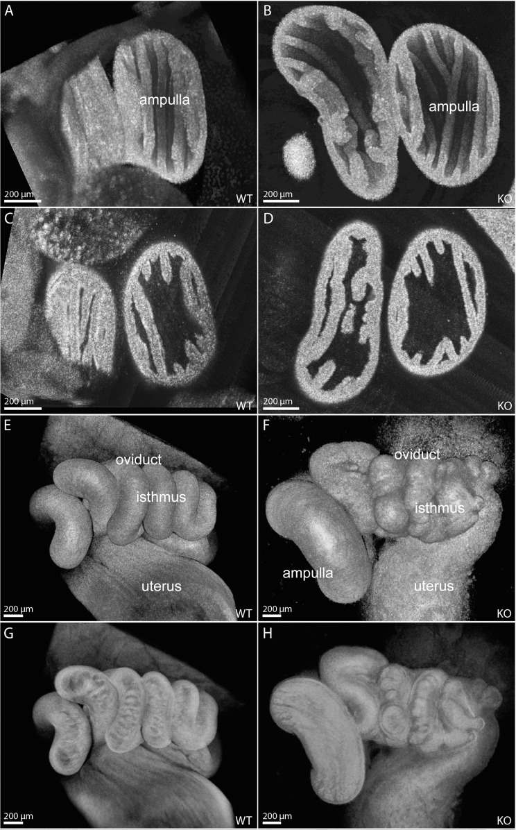
Structural oviduct abnormalities in 6-wk-old Smad1/5/4-Amhr2-cre KO mice visualized with OCT. Three-dimensional reconstructions visualizing the luminal surface of control (A) and Smad1/5/4-Amhr2-cre KO (B) female oviduct ampulla display similar levels of epithelial folding and density. Cross-sectional views through the reconstructions show comparable ampulla wall thicknesses in control (C) and Smad1/5/4-Amhr2-cre KO (D) female oviducts. Surface renderings of volumetric reconstructions of control (E) and Smad1/5/4-Amhr2-cre KO (F) female oviduct isthmus show an abnormal, uneven surface in the mutant. Semitransparent renderings of the corresponding data sets reveal abnormal disorganized luminal folding structures in the isthmus of the mutants (H) in contrast to those in controls (G).

