FIG. 5.
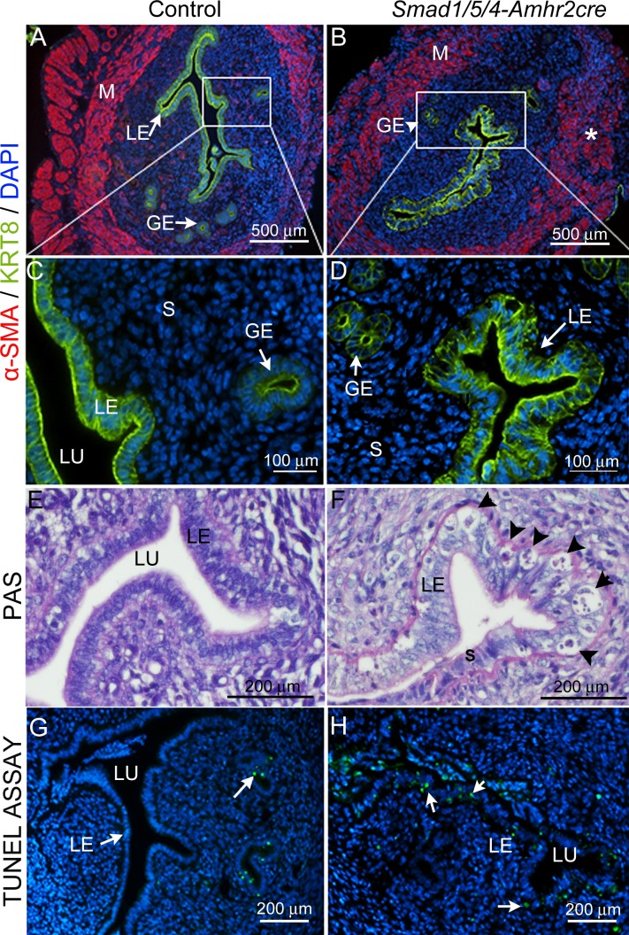
Loss of Smad1/5/4 disrupted smooth muscle development in uterus is shown and causes epithelial hyperplasia and apoptosis. Immunostaining of control (A and C) and Smad1/5/4-Amhr2-cre KO (B and D) in 6-wk-old uterus with the myometrium marker α-SMA (red) and the epithelial marker KRT8 (green). Insets in A and B are shown at 160X magnification for the luminal epithelium in C (control) and D (Smad1/5/4-Amhr2-cre KO). Compared to those in control, Smad1/5/4-Amhr2-cre KO uteri have disorganized myometrium (B*) and large vacuolated apoptotic cells in luminal epithelium (D and F). Six-wk-old sections of uteri were stained with PAS to show the histology of the uterine epithelium in control (E) and Smad1/5/4-Amhr2-cre knockout (F) mice. Vacuolated apoptotic cells in the luminal epithelium are indicated by arrowheads in F. G and H) TUNEL analysis for apoptotic cells (green) in uteri of 6-wk-old control (G) and Smad1/5/4-Amhr2-cre (H) KO mice. Nuclei are counterstained with DAPI (blue). Arrows indicate TUNEL-positive cells (green). GE, glandular epithelium; LE, luminal epithelium; M, myometrium; S, stroma; LU, lumen.

