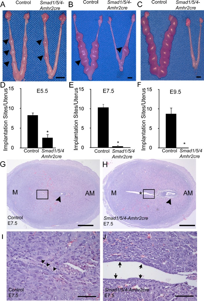
FIG. 6.
Smad1/5/4-Amhr2-cre mutants have implantation failure and defective embryo attachment. A–F) Mice were euthanized at E5.5, E7.5, and E9.5, and the numbers of implantation sites were counted in control and Smad1/5/4-Amhr2-cre KO mice. Arrowheads indicate implantation sites. Data are represented as means ± SEM; at least 3 mice were analyzed per genotype. Two-tailed unpaired Student t test was used to compare the control and Smad1/5/4-Amhr2-cre KO mice (*P < 0.05). G and H) H&E staining of control and Smad1/5/4-Amhr2-cre KO implantation sites at E7.5. G) Control embryo (arrowhead) with well-closed lumen and differentiated surrounding stroma. Insets from G and H are shown in I and J, respectively. H) Smad1/5/4-Amhr2-cre KO embryo (arrowhead) appeared to be degenerating, the uterus showed an *open lumen, and stromal cells have not fully differentiated. I and J) 125x magnification of insets shown in G and H, respectively. The luminal epithelium in control uteri has fully closed, and cells show signs of apoptosis (arrowheads). J). The luminal epithelium in knockout uteri remains visible (arrows). Bars = 5 mm (A–C), 400 μm (G and H), and 100 μm (I and J).

