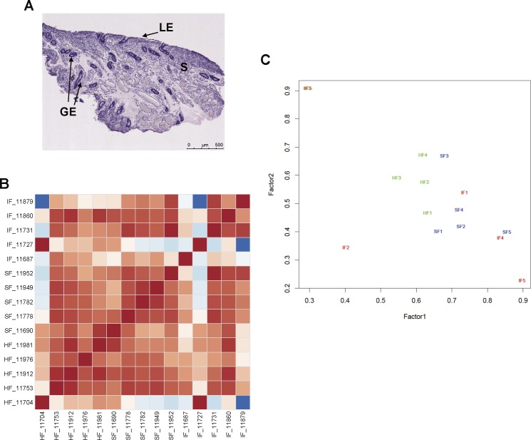
FIG. 4.
Endometrial biopsy histology and RNA sequencing analysis from Day 14 pregnant heifers. Fertility-classified heifers were synchronized to estrus and received two high-quality in vivo produced embryos on Day 7 postestrus. All heifers were nonsurgically flushed on Day 14 (7 days post-ET) to recover the conceptus. If a conceptus was present in the uterine flush, an endometrial biopsy was obtained from the uterine horn ipsilateral to the corpus luteum (CL). Total RNA was extracted from five biopsies of pregnant high fertile (HF), subfertile (SF), and infertile (IF) heifers and sequenced. Normalized and log2 transformed read count data were produced with edgeR-robust analysis. A) Histological analysis of a representative endometrial biopsy. All biopsies were predominantly composed of intercaruncular endometrium. Sections were stained with hematoxylin and eosin. LE, luminal epithelium; GE, glandular epithelium; S, stroma. Bar = 500 μm. B) Pairwise correlation (Pearson) analysis of gene expression levels between endometrial biopsy samples. Each column represents one sample and shows the correlation to all samples (including itself) with red for lowest (0) distance and blue for the highest observed distance. C) Multidimensional scaling plot. A maximum likelihood factor plot of gene expression variation among the samples. The major factors (factor 1 and factor 2) that explain the expression changes among the samples are plotted in the x- and y-axis, respectively. The individual samples (n = 15) representing the three fertility groups (HF, SF, and IF) are shown with different colors (green, blue, and red, respectively).

