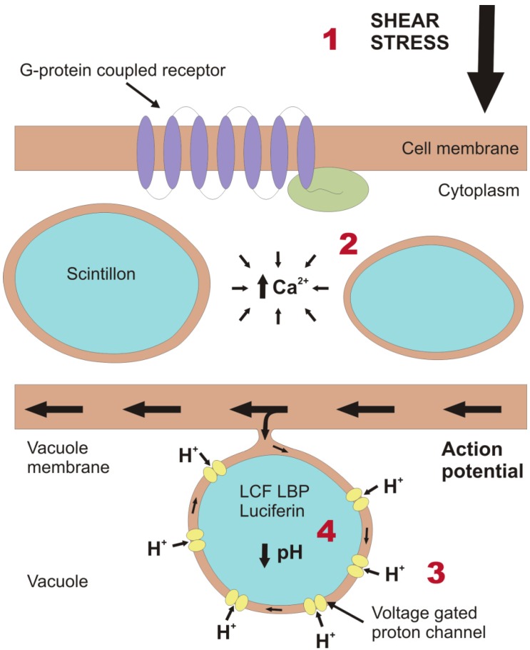
Figure 1.
Schematic representation of part of a dinoflagellate cell, depicting the cellular processes that take place to generate a bioluminescence flash. (1) Initially, a force is exerted on the outer cell membrane. Transduction involves the activation of a G-protein coupled receptor [22] (where G stands for GTP-binding), which typically consists of a receptor (purple) comprising seven transmembrane domains and the G-protein (green) on the inner side of the cell membrane. Subsequent to transmission of the mechanical signal from the outside to the inside of the cell, the signal must be translated to an electrical signal on the vacuole membrane. (2) This is achieved by increasing the concentration of Ca2+ in the cytoplasm, mainly from sources within the cell but also from the external medium [24]. This causes the vacuole membrane to depolarize and generate an action potential. In this illustration, one of the scintillons protrudes into the vacuole and is connected to the vacuole membrane. Thus, the action potential can travel across the membrane of the scintillon. (3) This results in the activation of voltage gated proton channels, which we here assume to be concentrated on the membrane of the scintillon rather than on the remaining vacuole membrane. (4) The influx of protons from the acidic vacuole rapidly lowers the pH of the scintillons and activates the bioluminescence chemistry. While some of the protons may be sequestered by the production of water as a by-product of bioluminescence, the fate of the rest of these protons and the mechanism by which the scintillons return to the original pH values are unknown.

