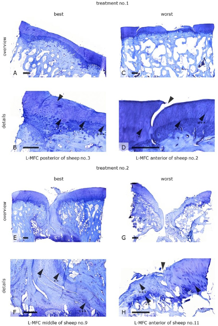
Figure 3.
Toluidine blue histology of tissue repair found in former defect sites (L-MFC) after being treated either with a biologic (Treatment No. 1) or a synthetic (Treatment No. 2) bilayer implant 19 months postoperatively. The figure shows examples of best versus worst specimens for both treatment groups. The defect sites were almost completely restored with a fibrous (F arrow*, H arrow***) and partly hyaline-like neocartilage with cells oriented in columns (B and D arrows***). The surface was either irregular (G) or smooth (A). The lateral integration was primarily good (B arrow). Lateral dehiscence was also found (D arrow). Tidemark formation (arrows**) indicated a sound cartilage-bone interface even in the worst specimens (H arrow**). Cell clusters at the boundaries respectively within the adjacent cartilage (B, D, H arrow*), a uniformly flat calcified layer, tidemark duplication, and occasional vascularization (F arrow***) indicated degenerative processes. These observations were variable and independent of implant design. Scale bars: 500 mm. L-MFC = left medial femoral condyle.

