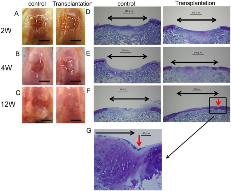
Figure 5.
Macroscopic and histological observation of the animals transplanted with cartilage-like cell-sheets. (A) At 2 weeks after transplantation, the surfaces of the defects were covered with a smooth, white, tissue layer. Control group, the defect was left empty. (B) At 4 weeks, the margins of the defects were unclear compared with control. (C) At 12 weeks, the repair tissue appeared similar to the surrounding articular cartilage. Control group, the defect was still left empty. Bar: 2 mm. Toluidine blue histology of the transplanted animals. Black arrow: cartilage defect. Red arrow: boundary between the transplant section and the normal cartilage. (D) At 2 weeks, the defects transplanted with cell-sheets were covered with a matrix that stained faintly with the metachromatic stain. Bar: 500 μm. (E) At 4 weeks, the defects were filled with stained tissue compared with control. Bar: 500 μm. (F) At 12 weeks, the defects transplanted with cell-sheets were filled with repair tissue compared with control. That repair tissue resembled hyaline cartilage. The repair tissue appeared continuous with the surrounding native cartilage tissue. Panel (G) is an enlarged view of the section in (F) identified by the red arrow. Bar: 40 μm.

