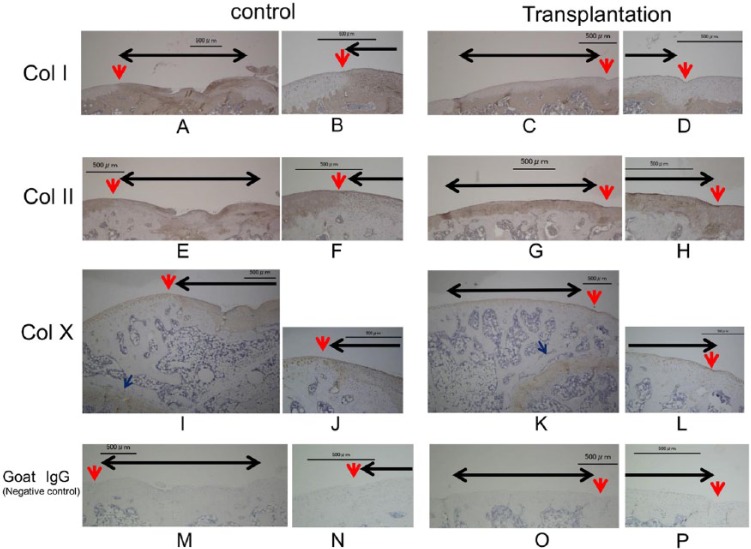
Figure 6.
Immunostaining for collagen types I (A, B, C, D), II (E, F, G, H), X (I, J, K, L), and negative control goat IgG (M, N, O, P) at 12 weeks after transplantation. B, D, F, H, J, L, N, P: magnified views of the sections in A, C, E, G, I, K, M, and O labeled with red arrows. Black arrow: cartilage defect. Red arrow: boundary between the transplant section and the normal cartilage. Blue arrow: growth plate. In the transplanted defects, staining was positive for collagen type II, but not collagen type I. Type X staining was weak compared with the growth plate and control defect. Bar: 500 μm.

