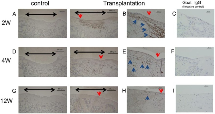
Figure 7.
Immunostaining for human vimentin (hVIM). Black arrow: cartilage defect. Blue arrow: positively stained cells. (A) At 2 weeks after transplant of the cartilage-like cell-sheets, the hVIM antibody showed a high level of human cell chimerism. (D) At 4 weeks, the amount of chimerism was decreased in the regenerated cartilage layer, but increased in the subchondral bone. (G) At 12 weeks, very low levels of chimerism were found. (B, E, H) Magnified views of the sections indicated by the red arrows in A, D, and G. (C, F, I) Goat IgG negative control sections. Bars: 500 μm (A, D, G); 40 μm (B, C, E, F, H, I).

