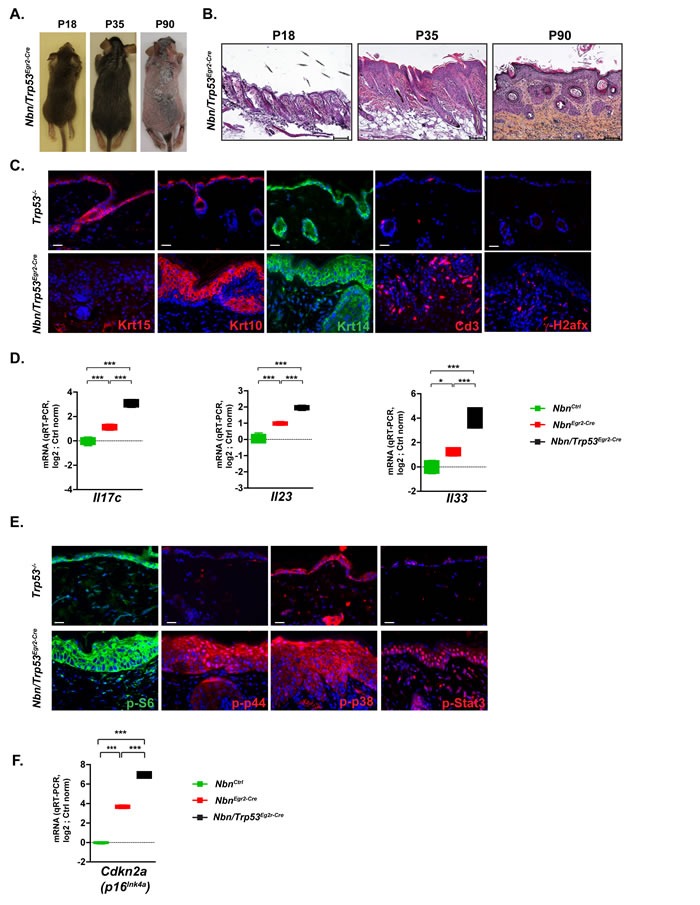
Figure 4. Trp53 inactivation triggers worsening of NbnEgr2-Cre phenotype.

A. The hair loss in Nbn/Trp53Egr2-Cre skins from P18 to P90 is similar to the NbnEgr2-Cre ones B. Histological analysis of Nbn/Trp53Egr2-Cre skins reveals progressive aggravation of NbnEgr2-Cre skins lesions by Trp53 inactivation at P90. Scale bar 100 μm. C. Characterization of Nbn/Trp53Egr2-Cre skins using Krt15, Krt10, Krt14, Cd3+ and γ-H2afx staining. Remark the lack of Krt15+ cells, the ectopic localization of Krt10- and Krt14-positive cells and the high number of DSBs revealed by γ-H2afx foci accumulation in Nbn/Trp53Egr2-Cre. Scale bar 20 μm.D. Dramatic increase of Il17c, Il23 and Il33 expression in Nbn/Trp53Egr2-Cre skins (NbnCtrl (N = 2), NbnEgr2-Cre (N = 2) and Nbn/Trp53Egr2-Cre (N = 2). E. Strong nuclear localization/activation of p-p44, p-p38 and p-Stat3 in Nbn/Trp53Egr2-Cre skins lesions. S6 phosphorylation occurs also in the basal keratinocytes layer. Scale bar 20 μm. F. Inactivation of Trp53 is associated with increase of Cdkn2a expression in Nbn/Trp53Egr2-Cre skins. NbnCtrl (N = 2), NbnEgr2-Cre (N = 2) and Nbn/Trp53Egr2-Cre (N = 2). ***: p < 0.001. Scale bar 20 μm.
