
Figure 2. ATGL down-regulation is associated with increased pro-inflammatory markers in C2C12 myoblasts.
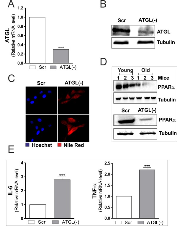
A. C2C12 cells were transfected with scramble (scr) or ATGL siRNA (ATGL(−)). Total RNA was isolated, and relative mRNA level of ATGL was analyzed by RT-qPCR. Data are expressed as means ± S.D. (n = 4, ***p < 0.0001 vs. scr cells). B. Twenty μg of total proteins were subjected to Western blot analysis of ATGL. Tubulin was used as loading control. C. Determination of TAGs content by Nile Red staining in scr and ATGL(−) myoblasts. D. Skeletal muscle of three young and three old mice was homogenized and 20 μg of total proteins were subjected to Western blot analysis of PPARα (upper panel). scr and ATGL(−) cells were lysed and 20 μg of total proteins were subjected to Western blot analysis of PPARα (bottom panel). Tubulin was used as loading control. All the immunoblots reported are from one experiment representative of three that gave similar results. E. Total RNA was isolated, and relative mRNA levels of IL-6 (left panel) and TNFα (right panel) were analyzed by RT-qPCR. Data are expressed as means ± S.D. (n = 4, ***p < 0.0001 vs. scr cells).