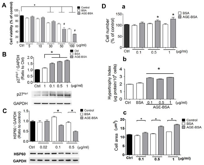
Figure 4. AGEs induce cell hypertrophy and decrease HSP60 expression in cultured β-cells.

Effects of AGEs on the cell viability A., p27Kip1 protein expression B., and HSP60 protein expression C. in RINm5f cells were shown. Cells were treated with AGE-BSA (5-100 μg/ml in A or 0.02-1 μg/ml in B and C) for 24 hours. Cell viability was determined by WST-8 assay. The protein expression was determined by Western blotting. Protein levels were quantified by densitometry and normalized by GAPDH levels. Moreover, effects of AGEs on the cell number (D-a), cell hypertrophy index (D-b), and cell area (D-c) of RINm5f cells were investigated. Cells were treated with AGE-BSA (0.1-1 μg/ml) for 24 hours. The viable cell number was determined by trypan blue exclusion assay. The cell hypertrophy index and cell diameter were measured as described under “Materials and Methods”. Data are presented as means ± SEM (n ≥ 5). *P < 0.05, versus BSA.
