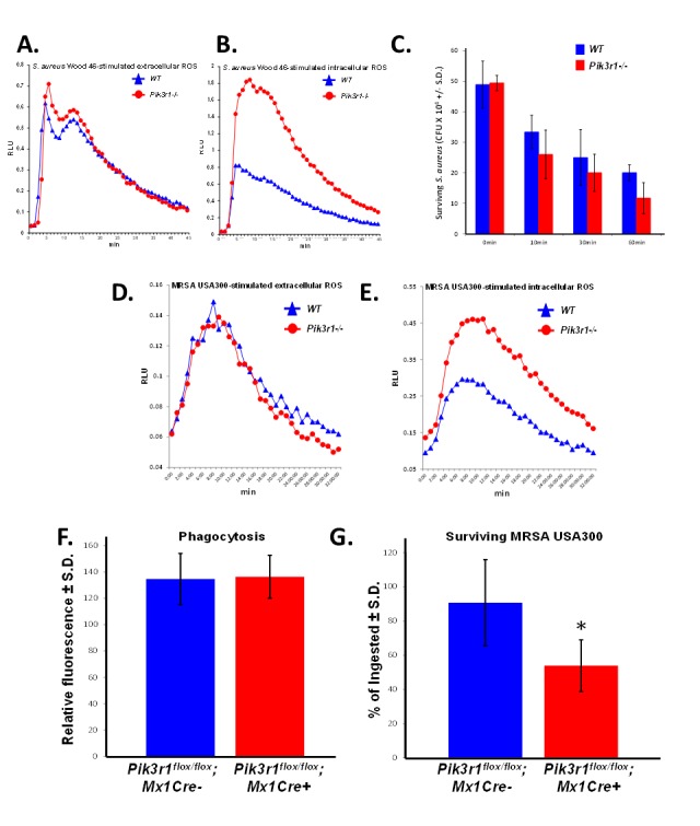
Figure 3. Pik3r1−/− neutrophils demonstrate superior killing of MSSA (Wood 46) and MRSA (USA300).

A. Extracellular and B. intracellular ROS production from WT and Pik3r1−/− fetal liver-derived neutrophils in response to serum-opsonized MSSA (Wood 46), representative of 2 independent experiments; C. WT and Pik3r1−/− fetal liver-derived neutrophil killing of MSSA (Wood 46) was measured by counting the number of surviving bacteria after 0, 10, 30, and 60min incubation, n = 3, p = 0.09 comparing Pik3r1−/− to WT at 60min, statistical analysis performed by unpaired, two-tailed Student's t-test; D. Extracellular and E. intracellular ROS production was measured in WT and Pik3r1−/− neutrophils stimulated with serum-opsonized MRSA (USA300), experiment performed on one occasion. F. Phagocytosis and G. killing of MRSA (USA300) by Pik3r1flox/flox; Mx1Cre− and Pik3r1flox/flox; Mx1Cre+ bone marrow neutrophils was measured by fluorescence remaining inside cells after washing and quenching extracellular fluorescence, n = 30, *p < 0.001 comparing Pik3r1flox/flox; Mx1Cre− to Pik3r1flox/flox; Mx1Cre+, statistical analysis by unpaired, two-tailed Student's t-test, experiment conducted on two independent occasions.
