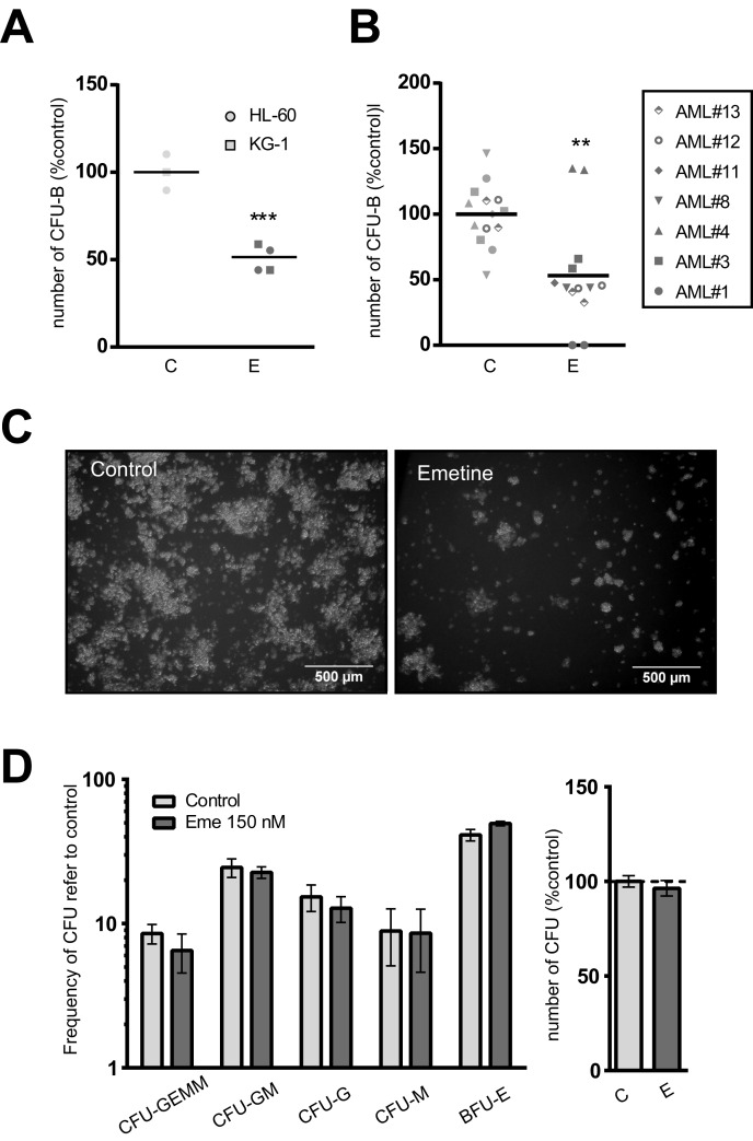
Figure 4. Emetine differentially reduced the clonogenic capacity of AML cells without affecting hematopoietic stem cell function.
A. AML cell lines or B. AML primary blasts were treated with 150 nM emetine (E, dark grey) or vehicle control (C, light grey) for 18 h. Colonies were screened at day 5 (HL-60), 7 (KG-1) or 14 (primary AML blasts). Each symbol represents a cell line or an AML primary sample. Results are normalized to control. C. Representative light microscope images of colonies in control- (left panel) and emetine-treated (right panel) HL-60 cells. D. Lineage-depleted umbilical cord blood cells were treated with emetine 150 nM (E, dark grey) or vehicle control (C, light grey) for 18 h. Colonies were screened at day 14 based on morphological and cellularity criteria. Left panel shows the frequency of each colony subtype relative to control, while right panel represents the total number of CFUs relative to control. Bars represent the mean value of two cord blood samples and error bars represent SEM. ** p < 0.01; *** p < 0.001.

