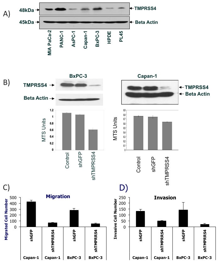
Figure 6. Verification of TMPRSS4 function in various PDAC cells.

A. TMPRSS4 protein expression across various PDAC cell lines and HPDE cells. Western Blot analysis of TMPRSS4 and beta actin. B. Cell viability of TMPRSS4 knockdown cells. Western Blot analysis of TMPRSS4 in whole cell lysates from BxPC-3 and Capan-1 cells 72 hours after shTMPRSS4 or shGFP infection (Top). Cell viability analysis of parental cells, shTMPRSS4 and shGFP cells using MTS assay (Bottom). C. + D. Knockdown of TMPRSS4 reduces migration and invasion of PDAC cells. Capan-1 cells or BxPC-3 cells stably transfected with shGFP-treated cells (control shRNA) or TMPRSS4 shRNA were placed in serum-free culture media and added into the upper compartment of a migration or invasion chamber. After 16 hours, cells in the upper chamber were removed and cells that had migrated or invaded through the pores of the membrane to the other side were fixed, stained, and counted. Cells in five different areas were quantified for migration and invasion studies. C, quantification of cells migrating through fibronectin-coated membranes. D, quantification of cells invading through Matrigel-coated membranes after 16-h incubation and 10% serum as chemoattractant.
