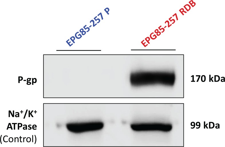
Figure 6. Quantification of P-gp expression in human gastric carcinoma cell lines.
Western blot analysis (10 μg protein/lane) was performed using JSB-1 monoclonal antibody directed against P-gp (170 kDa), to compare its expression in EPG85-257P cells and their MDR subline, EPG85-257RDB. Equal amounts of protein loading was confirmed using anti-KETTY antibody directed against the α–subunit of Na+/K+ ATPase (99 kDa). Protein concentration was determined by the Bradford protein assay.

