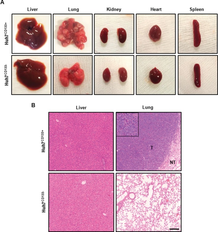
Figure 3. Gross photography of dissected organs and H & E staining from representative tumor from metastasis in vivo model.
(A) Representative images of dissected organs within the body after tail vein injection of irradiated Huh7CD133+ and Huh7CD133− cells. Multiple nodules were observed in both lung but none in Huh7CD133− injection group. In addition, no metastatic lesions were found in other organs except lung. (B) Representative H & E staining of the lung tissues in Huh7CD133+ injection group showed tumorous portion (T) and non-tumorous portion (NT). Tumors exhibited more aggressive invasion into surrounding lung tissue, undifferentiated and markedly pleomorphic tumor cells, suggesting poorly differentiated carcinoma. Scale bar, 200 μm.

